

Can MRIs Diagnose Autism? Will Autism Show On MRIs?

Autism is a neurodevelopmental disorder that affects communication, social interaction, and behavior.
It is estimated that 1 in 54 children in the United States has autism spectrum disorder (ASD). While there is no cure for autism, early diagnosis and intervention can improve outcomes for individuals with ASD.
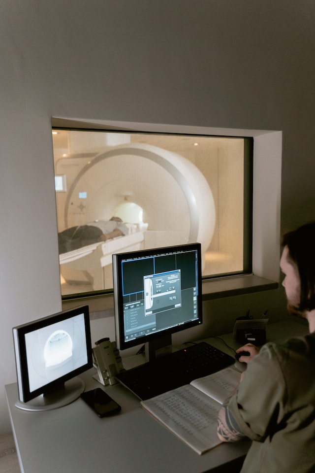
One question that often arises is whether autism can be detected through MRIs. MRIs, or magnetic resonance imaging, are a type of medical imaging that uses a magnetic field and radio waves to create detailed images of the body's internal structures.
While MRIs can detect structural abnormalities in the brain, such as tumors or lesions, they cannot diagnose autism.
Autism is a complex disorder that involves multiple brain regions and functions, and there is no single "autism center" in the brain that can be detected through MRI.
However, research has shown that there are differences in brain structure and function in individuals with autism compared to neurotypical individuals.
For example, some studies have found that individuals with autism have larger brains or differences in the size or connectivity of certain brain regions.
These differences are not specific to autism and can also be found in individuals with other neurodevelopmental disorders or even in neurotypical individuals. Therefore, while MRIs can provide valuable information about brain structure and function, they cannot be used as a diagnostic tool for autism.
Instead, the diagnosis of autism is based on behavioral and developmental assessments, such as the Autism Diagnostic Observation Schedule (ADOS) or the Autism Diagnostic Interview-Revised (ADI-R).
These assessments evaluate a child's communication, social interaction, and behavior, and are typically conducted by a trained professional, such as a psychologist or psychiatrist. Families with questions or those exploring support options often turn to evidence-based approaches like ABA Therapy to better understand their child’s unique strengths and needs.
What does autism look like in MRI?
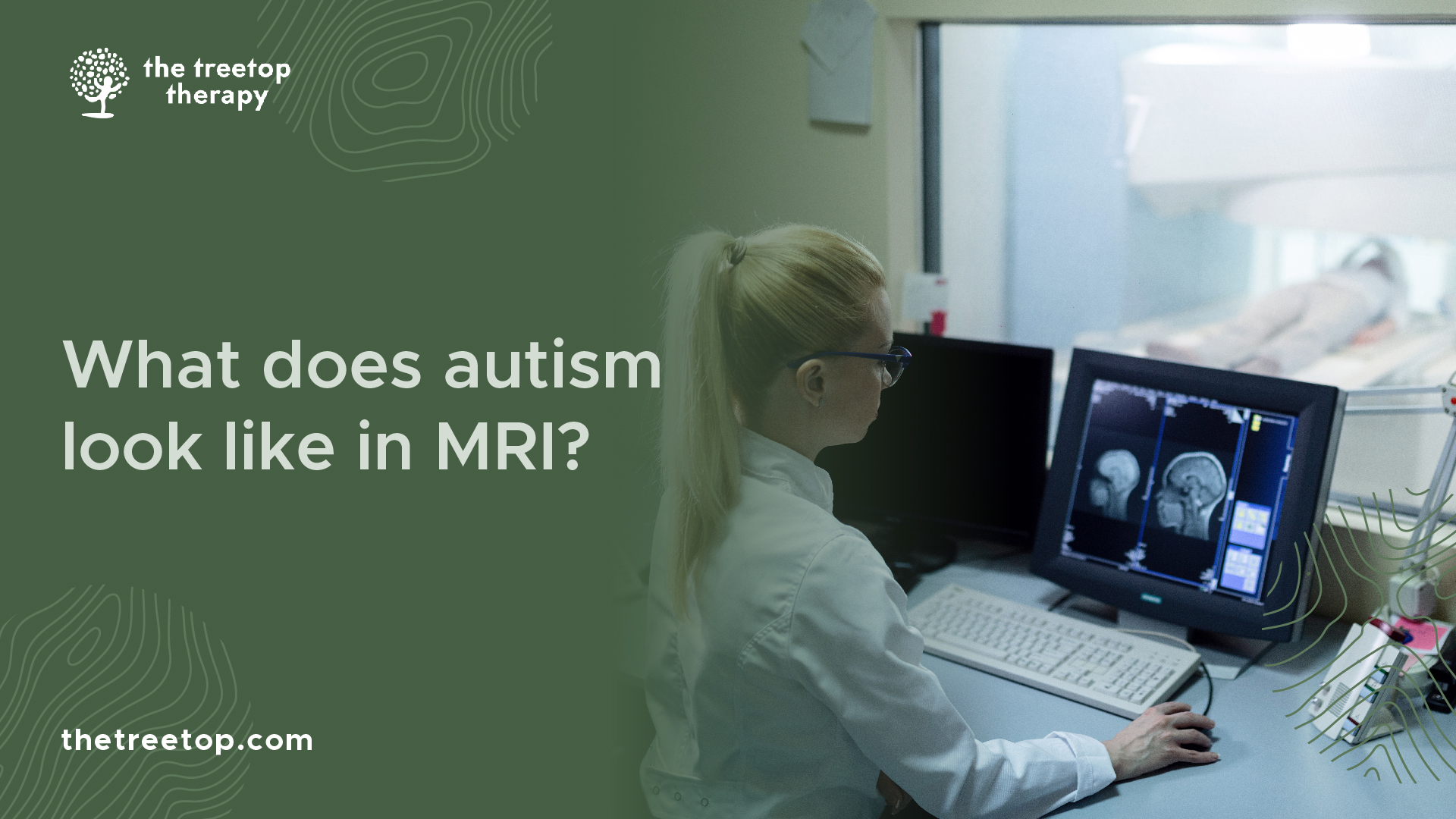
While MRIs cannot diagnose autism, they can provide valuable information about how autism affects the brain. For example, some studies have found that children with ASD tend to have more grey matter in certain areas of the brain, such as the prefrontal cortex and amygdala.
These areas are important for social communication and emotional regulation, which are often affected in individuals with autism.
Other studies have found differences in white matter connectivity between individuals with ASD and neurotypical individuals.
White matter is responsible for transmitting signals between different regions of the brain, so differences in connectivity may contribute to difficulties with communication and sensory processing that are common in individuals with ASD.
However, it's important to note that these findings are not consistent across all individuals with autism, and there is still much research to be done on how autism affects the brain.
Additionally, MRI findings alone cannot be used to diagnose or treat autism - behavioral assessments and interventions remain the gold standard for diagnosing and supporting individuals with this complex disorder.
Do autistic brains look different on MRI?
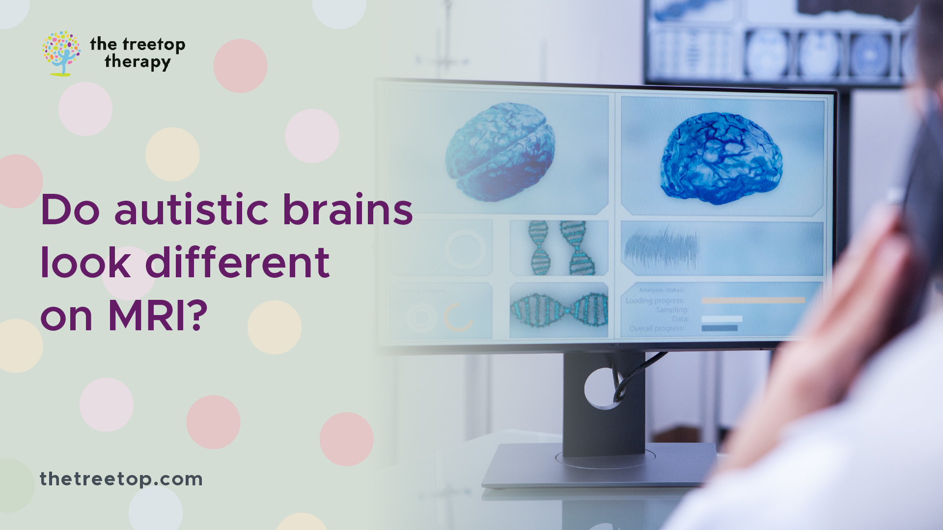
One common question about autism and MRIs is whether autistic brains look different on MRI scans.
While there is no single "autism center" in the brain that can be detected through MRI, research has shown that there are differences in brain structure and function in individuals with autism compared to neurotypical individuals.
Some studies have found that children with autism tend to have larger brains or differences in the size or connectivity of certain brain regions when compared to neurotypical individuals.
For instance, a study published in JAMA Psychiatry found that children with ASD had a larger amygdala than typically developing children. The amygdala is an almond-shaped region of the brain involved in processing emotions and social information.
Furthermore, another study published in Frontiers in Psychiatry found changes in the thickness of some cortical areas among autistic adults when compared with controls. Cortical areas play a key role in processing sensory information and integrating it into higher-order cognitive functions such as decision-making.
While these findings suggest that there are structural differences between autistic brains and neurotypical brains, they are not specific to autism and cannot be used alone for diagnosis.
Additionally, it's important to note that every individual with autism presents differently, so MRI results must always be interpreted cautiously by professionals trained in both neuroimaging and autism diagnosis.
Does autism always show up on a brain scan?
It's important to note that not all individuals with autism will have structural differences in their brains that can be detected through MRI. In fact, some individuals with autism may have brain scans that appear neurotypical.
This is because autism is a complex disorder that affects different individuals in different ways, and brain structure is just one aspect of the disorder.
Therefore, while MRIs can provide valuable information about how autism affects the brain, they are not a definitive diagnostic tool for the disorder.
Instead, behavioral assessments and evaluations remain the gold standard for diagnosing autism and developing appropriate interventions to support affected individuals.
What part of the brain is damaged in autism?
There is no single part of the brain that is damaged in autism. Rather, autism is a complex disorder that affects multiple brain regions and functions. However, research has identified several areas of the brain that may be involved in autism, including the prefrontal cortex, amygdala, and cerebellum.
The prefrontal cortex is located at the front of the brain and is responsible for executive functions such as decision-making, planning, and impulse control. Individuals with autism often have difficulties with these functions.
The amygdala is an almond-shaped structure located deep within the brain that plays a key role in processing emotions and social information.
Some studies have found differences in the size or connectivity of the amygdala in individuals with autism compared to neurotypical individuals.
The cerebellum is a region at the back of the brain that is involved in movement coordination and balance. Some studies have suggested that individuals with autism may have differences in cerebellar structure or function that contribute to difficulties with motor skills or sensory processing.
It's important to note that these are just a few examples of brain regions that may be affected by autism - there are many other regions and networks involved in this complex disorder.
Additionally, every individual with autism presents differently, so it's important not to generalize about how autism affects the brain without considering each person's unique profile.
In conclusion, while MRIs can provide valuable information about brain structure and function, they cannot diagnose autism.
The diagnosis of autism is based on behavioral and developmental assessments, and should be conducted by a trained professional.
Early diagnosis and intervention can improve outcomes for individuals with ASD, so it is important to seek evaluation if you have concerns about your child's development. To learn more about our team, values, and services, visit The Treetop ABA Therapy and see how personalized care can support long-term growth.